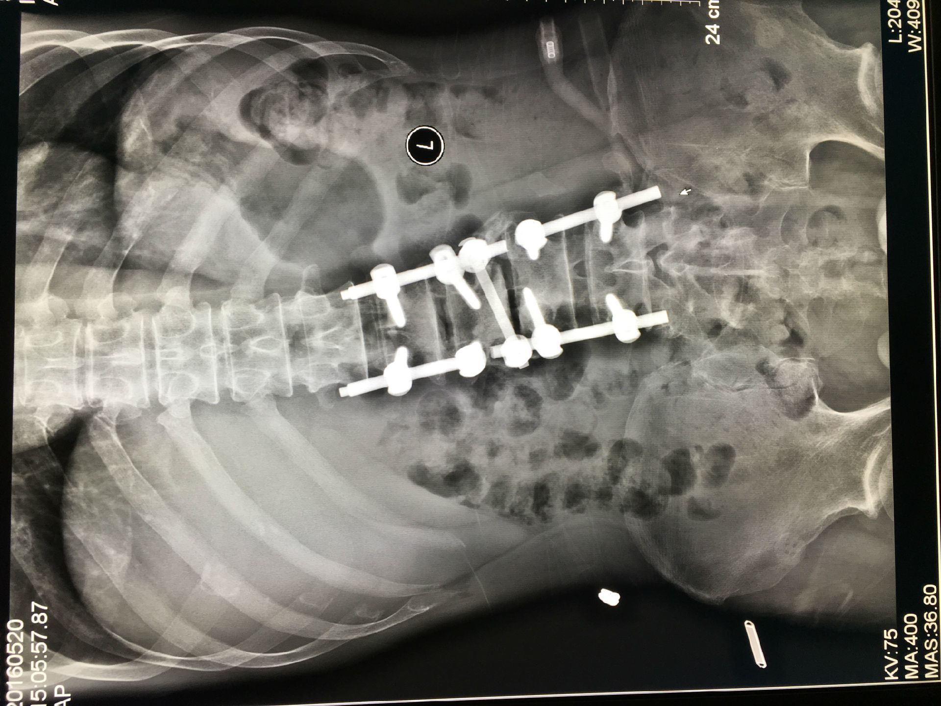
Spinal fusion is among the most commonly performed orthopedic surgeries
in the United States, with more than 400,000 patients undergoing the
procedure each year. Although most cases are successful, as many as 45
percent of patients experience complications after the operation, often
resulting from the bone's inability to tolerate the hardware surgeons
use to support the skeleton.
The most widely used technology to evaluate patients' bones before spine
fusion surgery is called dual x-ray absorptiometry, or DXA. DXA imaging
gives physicians a rough sense of the strength of a person's bone, but
it is not foolproof. In some cases, surgeons find that patients whose
DXA scans appear normal have bones that are so weak that the appliances
they use during spinal fusion are at risk of failure.
The HSS researchers hypothesized that a more sensitive measure of bone
quality could identify abnormalities in the skeleton that DXA does not
detect, and that these defects would be linked to postoperative
complications.

For the new study, the team led by Emily Stein, MD, an endocrinologist
and bone specialist, along with spine surgeons Han Jo Kim, MD, Matthew
Cunningham MD, PhD, and Frank Schwab, MD, turned to a cutting-edge
technique for assessing bone called high-resolution peripheral
quantitative computed tomography (HR-pQCT).
HR-pQCT can separately measure how much bone is in the outer (cortical)
and inner (trabecular) compartments and measure on a microscopic level
how the inner trabecular bone network is organized, including the
number, thickness and spacing between the parts of that network called
trabeculae. These measurements--known broadly as the
microarchitecture--may provide a much more robust assessment of skeletal
health than DXA, particularly in this population of patients who
frequently have changes in the area of the fusion which undermine the
utility of DXA.

The study included 54 men and women scheduled for spinal fusion at HSS
between December 2017 and December 2019. Patients underwent conventional
DXA scans, as well as HR-pQCT scans of the radius (forearm) and leg
bone (tibia).
Of the 54 patients in the study, 14 experienced complications within the
first six months of surgery, including broken rods, loosened bone
screws, fractures and abnormal bending of the spine. Although the number
of people in the study was small, the researchers found that those with
abnormalities on HR-pQCT were significantly more likely to experience
complications than those without such defects -- abnormalities that were
not evident on DXA imaging. These abnormalities involved lower bone
mineral density in the trabeculae (the spongy tissue in the center of
bones), fewer and thinner trabeculae, as well as thinner cortices.
"We used HR-pQCT to show for the first time that abnormalities in the
microscopic structure of bone are directly related to the development of
complications after fusion. Our study identified several abnormalities
in patients who had complications," Dr. Stein said.
For the moment, HR-pQCT is a research tool that's not widely available
to surgeons. "I think that it does provide a lot of additional
information that we're not getting from DXA. On DXA, the spine is almost
always going to look fine, which can be misleading," noted Dr. Stein.
Dr. Stein's group is expanding their study to include more patients.
"Spinal fusion surgeries are so invasive, the potential for
complications is high," she explained. "We want to have the most optimal
strategies for lowering complications in our patients, and that begins
with understanding who is at risk and why. In our future work with
additional patients, we hope to be able to define which features, or
group of features of the bone structure is most important in
contributing to surgical success. This will help us to devise the most
targeted treatment strategies for our patients."
About PUSM
Shenyang Pusman Medical Equipment Co., Ltd. was established in 2006. It is a professional enterprise integrating R & D, manufacturing and sales. It specializes in producing medical equipment according to national standards. Our company's main products: medical electric saw drill series; imported brushless motor series; domestic high-quality brushless motor series; medical electric drill pendulum saw integrated multi-functional system; hand, foot and ankle special system; pedicle screw screwing into special power; external Fixed needles are screwed into special power; external fixation series; bone cement; bone cement guns and other orthopedic surgical instruments.
Advantages of PUSM Power Tool
Integrated design, good sealing; integrated host provides you with efficient and stable output; reasonable distribution of the weight of the host from top to bottom, the user's hands are not tired; ergonomic design, comfortable feel; Suited sterilization box.

Contact:
Website: www.pusm-medical.com
Email: info@pusm-medical.com
Tel: +86-24-23519517
WhatsApp: +86 1399 8124 587